Playback speed
10 seconds
Video Player is loading.
Current Time 0:00
/
Duration 0:00
Loaded: 0%
0:00
Stream Type LIVE
Remaining Time -0:00
1x
- 0.5x
- 0.75x
- 1x, selected
- 1.25x
- 1.5x
- 1.75x
- 2x
- Chapters
- descriptions off, selected
- captions settings, opens captions settings dialog
- captions off, selected
This is a modal window.
Beginning of dialog window. Escape will cancel and close the window.
End of dialog window.
This is a modal window. This modal can be closed by pressing the Escape key or activating the close button.
0 views
June 17, 2012
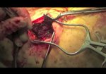
We aimed to present video illustration of scapulothoracic fusion technique in patients who have winging ...
read more ↘ scapula caused by neuromuscular disorders (mainly facioscapulohumeral dystrophy).
Patients with impaired shoulder function due to winging scapula were eligible for this procedure. Preoperatively meticulous physical examination and neurological consultation was made. Procerdure was contraindicated in patients with severe weakness of deltoid muscle or bad elbow function. They need to have at least 4/5 deltoid and, 3/5 biceps and triceps muscle strength to be included the study. Active shoulder ROM recorded while the scapula was free and while it was fixed manually. Shoulder motion, pain and satisfaction from the operation evaluated postoperatively.
Technique: Patients were operated in prone position. Longitudinal incision performed on the medial border of the scapula.Supraspinatus,infraspinatus and subscapularis muscles lifted subperiostally. Ribs and anterior surface of the scapula was deperiosted by burr. Cables(Dall-Miles cable grip system) passed beneath the ribs and through the holes drilled on the medial border of the scapula. Care was paid to prevent pleural injury. Autogenous graft from posterior iliac crest was enhanced with allograft and demineralized bone matrix. Graft mixture was located to the entire fusion area. Cables were tensioned and locked with specially
produced beads, starting from the relatively thicker inferior angle. Wound was closed over a drain.
Immobilization with arm sling was prescribed for at least 3 months.
Improvement in shoulder range of motion and functional scores were observed in follow-up.
↖ read less
Comments 0
Login to view comments.
Click here to Login