Playback speed
10 seconds
0 views
September 15, 2017
The purpose of this video is to demonstrate the surgical technique of closed reduction and ...
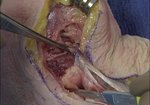
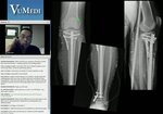
read more ↘ percutaneous pinning under fluoroscopic guidance of a displaced, pediatric extension type supracondylar humerus fracture. Intraoperative patient positioning, closed reduction maneuver, placement of 3 lateral smooth wires (0.062 inches) and postoperative splinting are demonstrated in this video. Pre-operative, intraoperative fluoroscopic and post-operative radiographic images are shown.
Title: Closed Reduction, Percutaneous Pinning of Supracondylar Humerus Fracture
Authors: Megan O’Connell, BS, David A. Fuller, MD, Cooper Medical School of Rowan University
Purpose: The purpose of this video is to demonstrate the surgical technique of closed reduction and percutaneous pinning under fluoroscopic guidance of a displaced, pediatric extension type supracondylar humerus fracture.
Methods: An 8 year old male with a displaced, extension type, extra-articular, supracondylar humerus fracture is shown undergoing closed reduction and percutaneous pinning of the fracture. Intraoperative patient positioning, closed reduction maneuver, placement of 3 lateral smooth wires (0.062 inches) and postoperative splinting are demonstrated in this video. Pre-operative, intraoperative fluoroscopic and post-operative radiographic images are shown.
Results: The video is 5min, 5 sec in duration.
Conclusion: Closed reduction and percutaneous pinning of a pediatric, supracondylar humerus fracture is demonstrated in this video.
↖ read less
Comments 3
Login to view comments.
Click here to Login
