Playback speed
10 seconds
Video Player is loading.
Current Time 0:00
/
Duration 0:00
Loaded: 0%
Stream Type LIVE
Remaining Time -0:00
1x
- 0.5x
- 0.75x
- 1x, selected
- 1.25x
- 1.5x
- 1.75x
- 2x
- Chapters
- descriptions off, selected
- captions settings, opens captions settings dialog
- captions off, selected
This is a modal window.
Beginning of dialog window. Escape will cancel and close the window.
End of dialog window.
This is a modal window. This modal can be closed by pressing the Escape key or activating the close button.
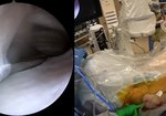
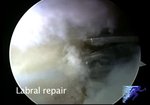
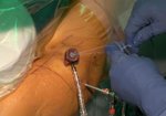
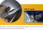
All Arthroscopic Single Row Repair Gluteus Medius Tendon Tear with Clinical Vignette and Preoperative Imaging.
0 views
April 20, 2011
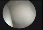
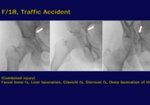
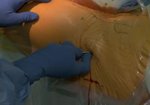
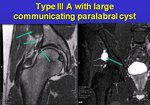
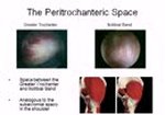
This video begins with a short history as explained by the patient. Preoperative MRI image ...
read more ↘ demonstrating gluteus medius tear is shown.
The arthroscopy, performed in the supine position using a 70 degree lens and three standard hip arthroscopy portals. Identification of the occult tear as visualized from the peritrochanteric space precedes tear mobility assessment, trochanteric footprint preparation, and suture anchor fixation to bone. A final video vignette demonstrating patient ambulation at 2 weeks closes the video.
<!--break-->
↖ read less
Comments 7
Login to view comments.
Click here to Login