Playback speed
10 seconds
Video Player is loading.
Current Time 0:00
/
Duration 0:00
Loaded: 0%
Stream Type LIVE
Remaining Time -0:00
1x
- 0.5x
- 0.75x
- 1x, selected
- 1.25x
- 1.5x
- 1.75x
- 2x
- Chapters
- descriptions off, selected
- captions settings, opens captions settings dialog
- captions off, selected
This is a modal window.
Beginning of dialog window. Escape will cancel and close the window.
End of dialog window.
This is a modal window. This modal can be closed by pressing the Escape key or activating the close button.
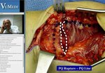
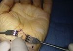
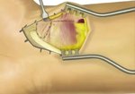
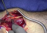
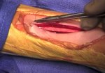
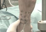
A Cadaveric Demonstration of the Volar (Henry) Approach to the Radius
0 views
July 30, 2024
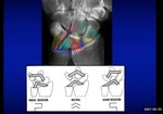
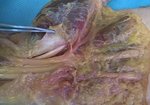
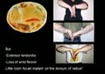
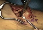
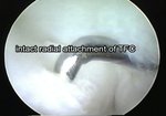
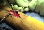
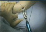
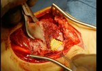
The volar approach to the radius is useful for a variety of indications such as ...
read more ↘ fracture fixation, compartment release, osteotomy, tumor resection, tendon repair, and nerve decompression. There are multiple structures at risk during this exposure, including the radial artery and its recurrent branch, posterior interosseous nerve, median nerve, and lateral antebrachial cutaneous nerve. In this video we demonstrate a safe approach to the volar radius, from the volar rim distally to the radiocapitellar joint proximally. We then perform an extended dissection of the forearm to show the course of the structures at risk and their proximity during exposure.
↖ read less
Comments 0
Login to view comments.
Click here to Login